So you want to run FACS on your microbiome samples…
By Cara Pardon
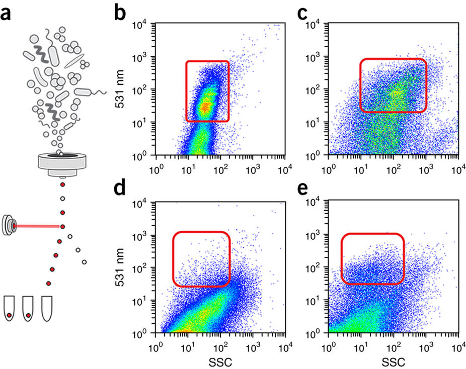
Although optimized for eukaryotic cell research, an increasingly common technique used in the microbiome field is Fluorescence Activated Cell Sorting (FACS). From sorting cells for single cell genomics to identifying the host range of a plasmid; from detecting metabolically active cells in an environmental sample to studying colitogenic-driving intestinal bacteria, FACS can be used in a multitude of applications to study microbial communities (Rinke et al. 2014, Klumper et al. 2015, Kalyuzhnaya et al. 2008, Hatzenpichler et al. 2016, Palm et al. 2014).
Are you interested in using FACS for your microbiome research? First, let me pose a question. What are the best practices when it comes to using appropriate controls for FACS microbial studies? From experience, I’ve learned that besides having negative controls (such as cells you know shouldn’t be carrying a marker, probe, or labeled secondary antibody) and positive controls (cells that are definitely labeled) to identify and verify the correct cells to sort, it may also be important to have controls of the various stages of sorting in the experiment. For example, what about comparing the unsorted cells, i.e. the total effluent from the FACS machine, with the original sample? Or comparing the sorted fraction to the unsorted bacterial population? Cells may lyse during the sort and therefore, not be ‘sorted’ at all. FACS is not perfect and can place environmental stress on cells, changing their shape or physiology during sorting. This may or may not be relevant in your data analysis. It is also important to compare your sorted fractions to a total sorted fraction to verify you are getting only and all the cells you believe you are sorting.
Nevertheless, there have been some promising results from studies employing FACS (and relevant controls) in novel ways.
Palm et al. used FACS with 16s sequencing to identify cells coated with IgA antibodies. FACS enriched for an IgA+ population of cells from an IBD patient that caused severe disease susceptibility, whereas IgA- cells did not. Kalyuzhnaya et al. used redox sensor green (RSG), a fluorescent dye, to detect metabolically active cells and enrich for environmental microbes in Lake Washington that could utilize methane. BONCAT, a method in which newly synthesized proteins are labeled, has been combined with FACS to probe the translational activity of microbes that oxidize methane in the ocean (Hatzenpichler et al. 2016). Lastly, I’ll point out the use of FACS to study the host range of fluorescently labeled plasmids isolated directly from soil (Klumper et al. 2015).
It can be difficult to verify results that rely on FACS of natural microbiomes. Therefore, I again will stress the importance of having all the necessary and useful controls. Klumper et al. for example, not only sorted fluorescently labeled transconjugants, but followed this with a second sorting, improving their enrichment, and further verified their samples with post-sorting selective plating and fluorescence microscopy. Plating is key since it is depends on something other than fluorescence. Similarly, Palm et al. used an orthogonal method—ELISA—to verify their sorted cells were IgA-coated. As a good control, Hatzenpichler et al. tested the specificity of their FACS gates by sorting different gates and analyzing them with fluorescence microscopy. Kalyuzhnaya et al. used a number of different controls. Firstly, they tested that cells that were metabolically labeled with RSG staining maintained their viability. They used a secondary dye propidium iodide, to confirm cells were dead, or rather dormant, and performed 16s PCR analysis. There are some controls that would improve overall confidence in FACS, such as using an unsorted fraction as a control. All of these verifications should be adopted wherever possible.
Although FACS can be used for a number of bacterial applications, results should always be taken with a grain of salt and critically analyzed for potential pitfalls. Don’t get me wrong. FACS provides great high through-put possibilities, especially for uncultivable populations. As more researchers continue to use FACS for an increasing number of microbiome applications, we will better understand its limitations, and develop better quality control methods.
Happy sorting!
Hatzenpichler, R. et al. (2016) Visualizing in situ translational activity for identifying and sorting slow-growing archaeal-bacterial consortia. Proc Natl Acad Sci USA 28: E4069-E4078.
Kalyuzhnaya, MG. et al. (2006) Fluorescence in situ hybridization-flow cytometry-cell sorting-based method for separation and enrichment of type I and type II methanotroph populations. Appl Environ Microbiol 72(6): 4293-301.
Klumper, U. et al. (2015). Broad host range plasmids can invade an unexpectedly diverse fraction of a soil bacterial community. ISME 9: 934-945.
Palm, NW. et al. (2014) Immunoglobulin A coating identifies colitogenic bacteria in inflammatory bowel disease. Cell (5)158: 1000-1010.
Rinke, C. et al. (2014) Obtaining genomes from uncultivated environmental microorganisms using FACS-based single-cell genomics. Nat Protoc 9(5): 1038-48.

Thank you for the excellent analysis! I would love you read any of your microbial cell sorting papers so I can learn from your protocols
Never mind, found your master’s thesis which is pretty informative :)